Title
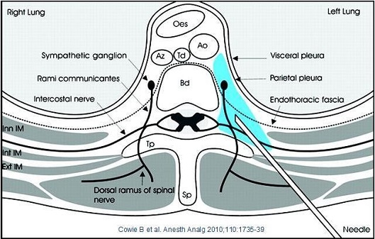
A cross-sectional view of the right lung and left lung which is located in the center of the image. The image is labeled with the names of the different parts of the lung including the sympathetic ganglion rami communicates intercostal nerve and endothoracic fascia.
The left lung is located on the left side of the body with the right side facing towards the viewer. The right lung is on the top left corner and the left lung on the bottom right corner.
There are several labels on the image including "Sympathetic ganglia" "Visceral pleura" and "Parietal pleura". These labels are likely used to indicate the location of the nerves in the neck and the location where the nerves are located. The diagram also shows the dorsal ramus of the spinal nerve which can be seen in the bottom left corner of the diagram.
- The image also includes a needle which may be used to connect the two parts together.
Type
Category
-
Date
2017
Collection
We encourage you to view the image in the context of its source document(s) and cite the source(s) when using these images. However, to cite just this image alone, click the “Cite This Image” button and then paste the copied text.


