OIDA Image Collection
Filters:
Reset 
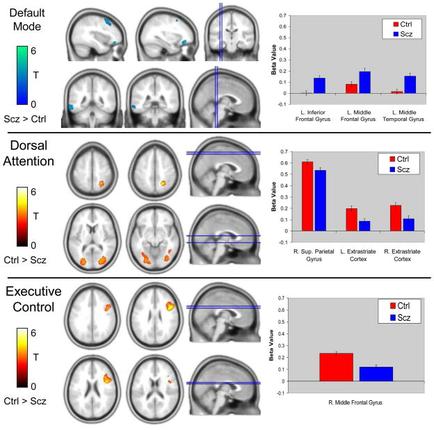
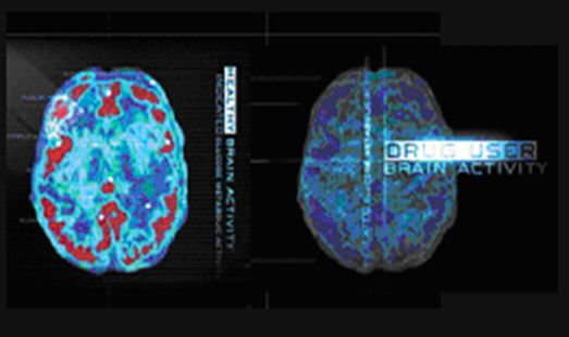
Two MRI scans of the brain one on the left side and the other on the right side. The scans are labeled "Drug User Brain Activity" and appear to be a comparison of the different areas of the human brain.<br /><br />The left scan shows the brain in red and blue colors while the right scan shows it in blue and green colors. The blue areas appear to have a more prominent area in the center of the image which is likely the location of the drug user brain activity. The image also has a label on the top right corner that reads "DRUG USER BRAIN ACTIVITY".<br /><br />Both scans are displayed on a black background and there is a text overlay on the image that explains the differences between the two scans.

Title

Brain activity imaging : Healthy brain vs. Drug user brain

Description Generated using AI

Two MRI scans of the brain one on the left side and the other on the right side. The scans are labeled "Drug User Brain Activity" and appear to be a comparison of the different areas of the human brain.

The left scan shows the brain in red and blue colors while the right scan shows it in blue and green colors. The blue areas appear to have a more prominent area in the center of the image which is likely the location of the drug user brain activity. The image also has a label on the top right corner that reads "DRUG USER BRAIN ACTIVITY".

Both scans are displayed on a black background and there is a text overlay on the image that explains the differences between the two scans.

Category

We encourage you to view the image in the context of its source document(s) and cite the source(s) when using these images. However, to cite just this image alone, click the “Cite This Image” button and then paste the copied text.
Information copied to your clipboard:

"Brain activity imaging : Healthy brain vs. Drug user brain." OIDA Image Collection. Available at https://images.oida-resources.jhu.edu/032b7f3f-2eef-...