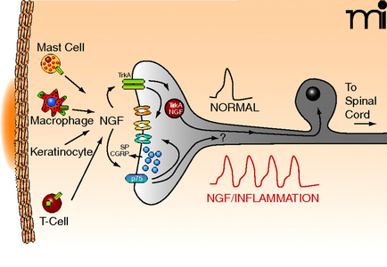
A diagram that shows the structure of a mast cell and its functions. On the left side of the image there is a cross-section of the mast cell which is a type of cell that is responsible for the formation of a T-cell. On the right side there are two types of cells one labeled "Macrophage NGF" and the other labeled "Keratinocyte". The T-Cell is a protein that binds to the NGF (NGF) in the cell membrane. The NGF is a neurotransmitter that helps to regulate the flow of neurotransmitters in the body. In the center of the diagram we can see a normal cell with a spiral cord attached to it. The spiral cord is used to connect the T-cells to the spinal cord which helps to stimulate the cell's nervous system. This helps to reduce the risk of inflammation and improve the overall health of the body's immune system. The image also shows a red line graph that shows a decrease in NGF/inflammation which can be seen in the bottom right corner.
Description 
Type
Category
Source 1 of 3
-
Date
2018
Collection
-
Date
2018
Collection
-
Date
2018
Collection
We encourage you to view the image in the context of its source document(s) and cite the source(s) when using these images. However, to cite just this image alone, click the “Cite This Image” button and then paste the copied text.