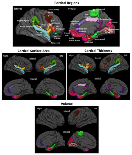
A collage of six different views of the human brain each showing a different area of the brain. The top left view shows the lateral and cortical regions of the head with the lateral area on the left side of the image and the cortical surface area in the center. The lateral area is labeled with the names of the different areas while the cortical areas are labeled with their respective names. In the top right view there is a diagram of the cerebral cortex which is a type of brain that shows the location of the cerebellum in the brain and the surrounding area. The cerebral cortex is colored in different shades of green blue red and purple representing the different regions. The medial area is colored with a red and green color scheme representing a different color scheme. The left side shows the left and right sides of the cortex with a green and red color scheme representing the medial and medial areas and a purple and green area representing a medial area. - The top middle view shows a diagram that shows that the cerebral area is divided into two sections one labeled "Cortical Surface Area" and the other labeled "Volume". The right side shows that there are two sections labeled "Lateral" and "Cortex Thickness". Overall the image shows the different types of cortical regions in the head and how they interact with each other.
Description 
Type
Category
Source 1 of 6
-
Date
2015
Collection
-
Date
2015
Collection
-
Date
2015
Collection
-
Date
2015
Collection
-
Date
2015
Collection
-
Date
2015
Collection
We encourage you to view the image in the context of its source document(s) and cite the source(s) when using these images. However, to cite just this image alone, click the “Cite This Image” button and then paste the copied text.