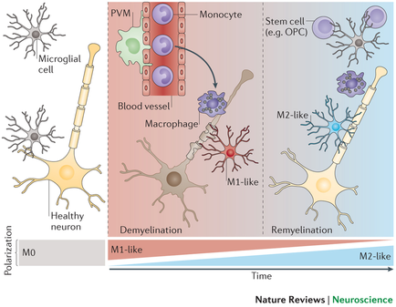
A diagram that shows the different types of neurons in the brain. The diagram is divided into three sections. The top section shows a microbial cell which is a type of nerve cell that is responsible for regulating the flow of blood vessels in the nervous system. The cell is represented by a red circle with the letters "PVM" and "Monocyte" inside it. The red circle represents the blood vessel while the blue circle represents a stem cell (e.g. OPC) and the purple circle represents stem cells. In the center of the diagram there is a blood vessel with a macrophage which helps to regulate the blood flow. The blood vessel is connected to the stem cell by a membrane. The membrane is also connected to a membrane that helps to store the blood vessels. The image also shows a healthy neuron which can be seen in the bottom left corner of the image. The neuron is shown as a yellowish-orange color and is labeled as "M1-like". On the right side of the illustration there are two neurons one labeled "M2-like" and the other labeled "Remyelination". These neurons are shown as yellowish orange and are connected by a blue line. The diagram also shows the time and nature reviews for neuroscience.
Description 
Type
Category
-
Date
2015
Collection
We encourage you to view the image in the context of its source document(s) and cite the source(s) when using these images. However, to cite just this image alone, click the “Cite This Image” button and then paste the copied text.