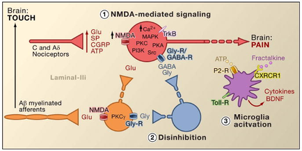
A diagram that shows the process of NMDDA-mediated signaling in the brain. It shows the different types of neurons and their functions. The diagram is divided into three sections. The first section is labeled "Brain: Touch" and shows the interaction between the brain and the receptors. The neuron on the left side of the image is labeled as "C and A6 acceptors" and the receptor on the right side is labelled as "Laminal-III". The receptor is represented by a red circle with the letters "NMDDA" and "MPK" written on it. The receptors are represented by two orange circles one labeled "glue" and another labeled "PKG" which are the receptors that are responsible for the signaling of the NMDAs. The red circle represents the receptors in the receptor while the orange circle represents a receptor that binds to the receptor. The blue circle represents an ATP (ATP) receptor which is responsible for signaling the receptor to the receptors and the purple circle represents cytosine's (BCNF). There is also a diagram on the bottom right corner that shows how the receptor interacts with the receptor and how it interacts with other receptors such as "Toll-R" "Microglia activation" and "Fractalkine". Overall the diagram shows how NMDAA-mediated signals can be used to signal the presence of a receptor in a brain which can be controlled by the receptors and receptors.
Description 
Type
Category
Source 1 of 3
-
Date
2014
Collection
-
Date
2014
Collection
-
Date
2014
Collection
We encourage you to view the image in the context of its source document(s) and cite the source(s) when using these images. However, to cite just this image alone, click the “Cite This Image” button and then paste the copied text.