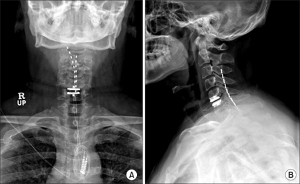
A collage of two X-ray images of a human neck and upper body. The left side of the image shows the neck and neck area while the right side shows the back of the neck. The image is labeled "RUP" and "A" and is labeled as "B". In the image there is a cross-section of the human neck showing the vertebrae and the spinal cord. The spinal cord is visible in both images with the RUP on the left and the B on the right. The X-rays appear to be in good condition with no visible signs of wear or damage. The image also shows a small amount of blood or fluid in the neck which is likely due to the presence of a large amount of fluid. The RUP is located in the center of the spine and it appears to be attached to the neck with a metal rod. The metal rod is likely a medical device possibly a surgical instrument and is likely used to measure the size and shape of the vertebral column. The background of both images is black making the white lines stand out.
Description 
Type
Category
-
Date
2016
Collection
We encourage you to view the image in the context of its source document(s) and cite the source(s) when using these images. However, to cite just this image alone, click the “Cite This Image” button and then paste the copied text.


![A screenshot of the Formulary update page of the Xartemis™ XR (oxycodone HCI and acetaminophen Extended-Release Tablets (CII) website. The page has a white background with a green and purple color scheme. The header reads "Formulary update" in bold purple font. Below the header there is a text box that reads "XARTEMIS XR is available unrestricted on Tier 3 for State of Connecticut EMP (CT) Commercial Patients. No PA/Step therapy requirement. Patients will have an average co-pay of [S]." <br /><br />At the bottom of the page there are four buttons each reading "Formulary Update". The website appears to be a mock-up and it appears that some placeholder text exists between brackets.<br /><br />The page also has a navigation bar at the bottom of the page which shows that the "Formulary Updates" section is selected.](https://oida-resources-images.azureedge.net/public/full/c4b34ebc-dbfb-4b3f-b1f4-387298601786.jpeg)