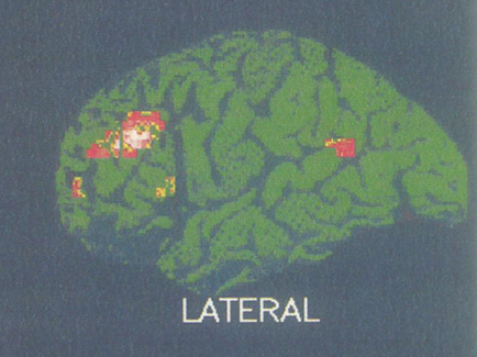
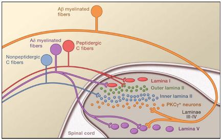
A diagram that shows the structure of a spinal cord and its functions. The diagram is divided into three sections. The top section is labeled "A8 unmyelinated fibers" and shows the different types of fibers that make up the spinal cord. The fibers are represented by different colors - blue purple and orange. The blue and purple fibers are labeled as "nonpeptidegic fibers" the purple and orange fibers are labelled as "PEPTIDERGIC C fibers" and the orange fiber is labeled with "Lamina I". In the center of the diagram there is a network of interconnected lines that represent the different fibers. The lines are connected by arrows indicating the flow of information between the fibers. On the right side of the image there are several smaller circles representing the different functions of the fibers such as "outer lamina II" "inner lamina III" "pkwy" and "Laminae III-IV". The circles are colored in different shades of blue green and purple representing different regions of the body. The arrows indicate the direction of the flow from the top left corner to the bottom right corner. The background is a light beige color which makes the colors of the lines stand out.
Description 
Type
Category
Source 1 of 4
-
Date
2014
Collection
-
Date
2014
Collection
-
Date
2015
Collection
-
Date
2014
Collection
We encourage you to view the image in the context of its source document(s) and cite the source(s) when using these images. However, to cite just this image alone, click the “Cite This Image” button and then paste the copied text.