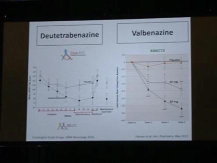
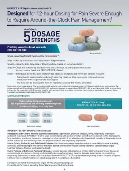
A diagram that shows the structure of a protein molecule. It consists of two parts - a and b. The top part of the diagram shows the protein molecule HVACC which is a type of protein that binds to the vac (HVACC) molecule. The molecule is represented by a blue circle with a green arrow pointing to it. The arrow is connected to the molecule with a red line. In the diagram there are two proteins AKT (AKT) and GSK3B which are responsible for the formation of the protein in the molecule. AKT is a protein that is responsible for binding to the protein while GSK2B is a compound that binds the protein to the other proteins. The molecules are represented as green and red arrows representing the proteins that bind to each other. The red arrows represent the proteins in the molecules while the green arrows represent proteins in their respective proteins such as ATP (ATP) and CAMP (CAMP). At the bottom of the image there is a label that reads "Nature Reviews | Neuroscience". This label indicates that the image is related to nature reviews and neuroscience.
Description 
Type
Category
-
Date
2013
Collection
We encourage you to view the image in the context of its source document(s) and cite the source(s) when using these images. However, to cite just this image alone, click the “Cite This Image” button and then paste the copied text.