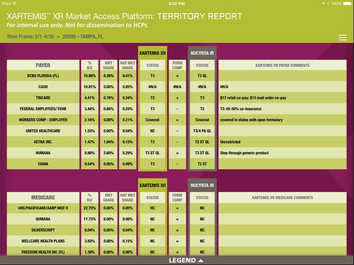
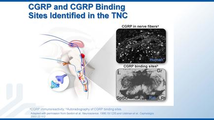
A slide from a presentation about carp and carr binding sites identified in the TNC. The slide has a blue background with white text. On the left side of the slide there is an illustration of a human brain with a red and blue neuron in the center. The neuron is surrounded by a white circle with a blue line connecting it to the brain. On the right side there are two images - one is a black and white image of a brain scan and the other is a close-up of the brain scan. The brain scan shows the brain with the red and white neurons while the blue and red neurons represent the neurons. The image also has text that explains that the image is related to the carp binding sites which are used to bind nerve fibers in the brain to the spinal cord. The text also mentions that the images are related to corps which is a type of nerve fibers that are responsible for binding nerve fibers.
Description 
Type
Category
-
Date
2014
Collection
We encourage you to view the image in the context of its source document(s) and cite the source(s) when using these images. However, to cite just this image alone, click the “Cite This Image” button and then paste the copied text.