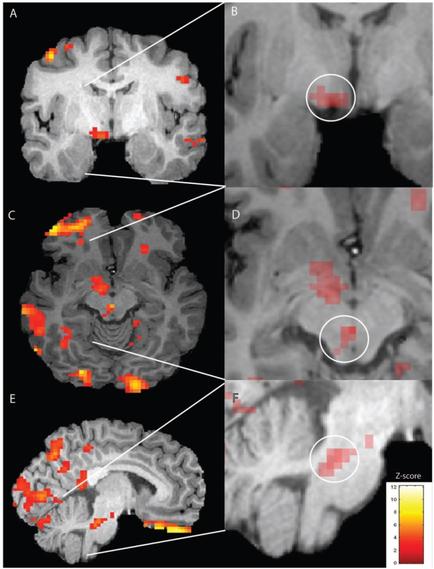
A collage of four MRI scans of the human brain. The scans are arranged in a grid-like pattern with each scan showing a different view of the brain. The top left scan shows the left side of the image with the top right scan showing the brain in black and white. The brain is shown in red and orange with a red dot in the center. The red dot appears to be a representation of the cerebellum while the orange dot is a representation from the brainstorm. In the top center scan there is a white circle with a black outline which is likely the location of the tumor. The white circle is likely a tumor as indicated by the red dot. The image also shows a red arrow pointing to the tumor indicating that the tumor is located in the right side of this scan. - The bottom left scan is a close-up of the left brain showing the different areas of the skull including the cerebrum cerebellums and brainstorm as well as the surrounding area. The bottom right scan is an MRI scan of the right brain which shows the same tumor but with a different color scheme. The colors in the image are red orange and yellow which may indicate the presence of a tumor in the skull.
Description 
Type
Category
Source 1 of 2
-
Date
2018
Collection
-
Date
2018
Collection
We encourage you to view the image in the context of its source document(s) and cite the source(s) when using these images. However, to cite just this image alone, click the “Cite This Image” button and then paste the copied text.