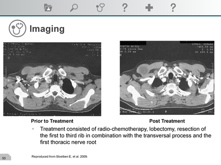
A screenshot of an MRI scan of a patient's head and neck. The scan is divided into two sections one on the left side and the other on the right side. The left side of the image shows the head of the patient with a large tumor in the center. The tumor appears to be in the middle of the head with a small amount of blood vessels surrounding it. The blood vessels are white and appear to be inflamed. The image is labeled "Prior to Treatment" and "Post Treatment". Below the image there is a text that explains that the patient is treated with radio-chemotherapy lobectomy resection of the first to third rib in combination with the transversal process and the first thoracic nerve root. The text also mentions that the treatment was performed from St. Joseph E. et al. 2009. At the top of the screenshot there are several icons including a magnifying glass a question mark and a calculator.
Description 
Type
Category
-
Date
2015
Collection
We encourage you to view the image in the context of its source document(s) and cite the source(s) when using these images. However, to cite just this image alone, click the “Cite This Image” button and then paste the copied text.