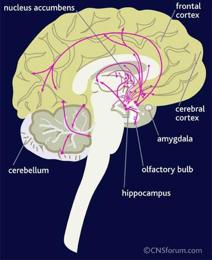
A cross-section of the brain showing the different parts of the human brain. The brain is shown in a light yellow color with the nucleus accumbent frontal cortex cerebral cortex amygdala olfactory bulb and hippocampus visible. The brain is divided into different sections including the cerebellum the frontal cortex (frontal cortex) and the hippocampus (hippocampus). The frontal cortex is located on the left side of the image while the cerebral cortex is on the right side. The amygdala is located in the center of the head with a pink line connecting it to the frontal lobe. The hippocampus is located at the bottom right corner of the diagram. There are also several red lines connecting the various parts of this brain which appear to be nerves and arteries. These lines are likely representing the nerves and nerves that make up the brain's structure. The image is labeled with the names of the different types of nerves and their functions.
Description 
Type
Category
-
Date
2012
Collection
We encourage you to view the image in the context of its source document(s) and cite the source(s) when using these images. However, to cite just this image alone, click the “Cite This Image” button and then paste the copied text.