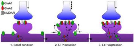
A diagram that shows the process of making a LTP expression in a cell. It consists of three stages of the process: 1. Basal condition 2. LTP induction 3. LTTP expression The first stage is shown in the top left corner of the image. It shows a blue flask with a red label that reads "GluA1" and "GluA2" on the top right corner. The flask is connected to a blue cylinder with a green label that says "NMDAR". In the middle stage there is a purple cylinder with red and green labels that read "Ca2". The label also mentions that the LTP is a type of protein that binds to the cell membrane. There are also two smaller blue vases in the image one on the left side of the diagram and the other on the right side. The vases are connected to each other by a series of red and yellow labels. The red labels indicate that the protein binds to a specific protein while the green labels indicate the different types of proteins that are attached to the membrane. The blue labels also mention that the proteins are responsible for the expression of the protein in the cell which is represented by the red labels on the labels.
Description 
Type
Category
Source 1 of 3
-
Date
2015
Collection
-
Date
2014
Collection
-
Date
2014
Collection
We encourage you to view the image in the context of its source document(s) and cite the source(s) when using these images. However, to cite just this image alone, click the “Cite This Image” button and then paste the copied text.