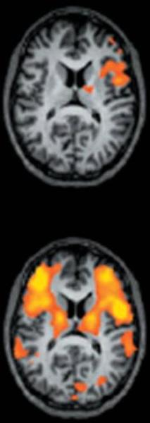
A CT scan of the brain which is a type of MRI scan that shows the brain in two different views. The scan is circular in shape and appears to be a cross-sectional view of the human brain. In the top view the brain is shown in black and white with the top half of the image being the largest and the bottom half being the smallest. The brain is divided into two sections with a central area in the center and a smaller area on the left side. The central area is filled with orange and yellow areas which appear to be the areas where the brainstorm is located. The orange areas are likely the areas that are affected by the brain activity while the yellow areas are areas where it is affected by a brain activity. - The image is taken from a top-down perspective and the background is black making the orange areas stand out. The image appears to have been taken at night as there are no other objects in the image.
Description 
Type
Category
-
Date
2012
Collection
We encourage you to view the image in the context of its source document(s) and cite the source(s) when using these images. However, to cite just this image alone, click the “Cite This Image” button and then paste the copied text.