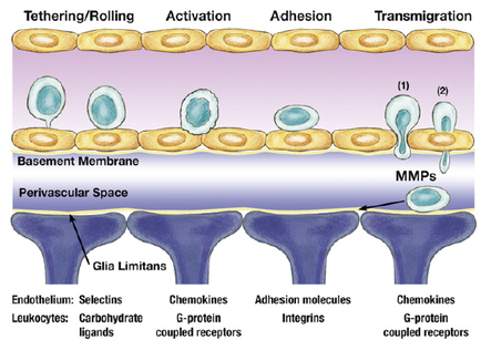
A cross-section of a cell membrane showing the structure of the membrane. The membrane is divided into two sections with the top section showing the tethering/rolling process and the bottom section showing a transmigration process. The membrane is made up of a basement membrane which is a type of membrane that separates the membrane from the rest of the body. The top section shows the activation and adhesion of the cells which are responsible for the transfer of the transfer. The activation is represented by a blue circle while the adhesion is shown by a yellow circle representing the transfer process. There are also several other components in the image including the membrane membrane the perivascular space and the endothelium. These components are labeled with their names such as "Gia limits" "Chemokines" "Adhesion molecules" "integrity" and "G-protein coupled receptors". These components represent the different types of receptors that are used in the membrane to transfer the transfer from one cell to another. The image also shows the endothelialium which helps to select the leukocytes carbohydrates and ligands of the cell membrane.
Description 
Type
Category
-
Date
2015
Collection
We encourage you to view the image in the context of its source document(s) and cite the source(s) when using these images. However, to cite just this image alone, click the “Cite This Image” button and then paste the copied text.