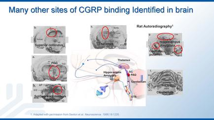
A diagram that shows the different sites of carp binding in the brain. It is divided into six sections each representing a different site. The first section on the top left shows a super-collicularis (super-colloidal) with a red circle in the center. The second section shows a rat autoradiographs (Rat Autoradiograph) with the text "Hippocampus amyloid" and "Cerebellum" written on it. The third section shows an illustration of the brain with a blue arrow pointing to the left side of the image. The fourth section shows the brainstorm with a white arrow pointing towards the right side. The fifth section shows two red circles one labeled "PAG" and the other labeled "CEREMBULUM". The sixth section shows three red circles with text that reads "Adapted with permission from Sexton et al. Neurosciences 1986-1995". Overall the image is an illustration that shows how the corps binding in a brain can be used to identify different sites.
Description 
Type
Category
-
Date
2014
Collection
We encourage you to view the image in the context of its source document(s) and cite the source(s) when using these images. However, to cite just this image alone, click the “Cite This Image” button and then paste the copied text.