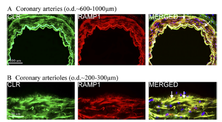
A close-up of a tissue sample which appears to be a microscopic view of a human body. The tissue is divided into two sections with the top section showing the internal structure of the tissue and the bottom section showing a small yellow-colored cell. The cell is located in the center of the image and is surrounded by several red-colored cells. The image is accompanied by text that reads "Reimbursement Assessment" and "December 20 2010" at the bottom. The CENDYL logo is also visible in the bottom right corner.
Description 
Type
Category
-
Date
2012
Collection
We encourage you to view the image in the context of its source document(s) and cite the source(s) when using these images. However, to cite just this image alone, click the “Cite This Image” button and then paste the copied text.