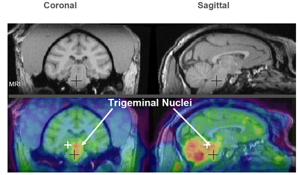
A collage of three MRI scans of the brain labeled as "Coronal" "Sagittal" and "Trigeminal Nuclei". The scans are arranged in a grid-like pattern with each scan showing a different view of the human brain. The first scan on the top left shows the normal brain with the left side of the image showing the cerebellum and the right side showing the trigeminal nerve. The brain is shown in black and white while the scan on top right shows the sagittal nerve which is a type of nerve that connects the brain to the spinal cord. The image also shows a cross-sectional view of a tumor in the brain. The tumor appears to be in the center of the head with a red arrow pointing to the left and a green arrow pointing towards the right. The red arrow is likely indicating the location of the tumor which may be a tumor or a tumor that may have been affected by the tumor.
Description 
Type
Category
Source 1 of 2
-
Date
2014
Collection
-
Date
2014
Collection
We encourage you to view the image in the context of its source document(s) and cite the source(s) when using these images. However, to cite just this image alone, click the “Cite This Image” button and then paste the copied text.