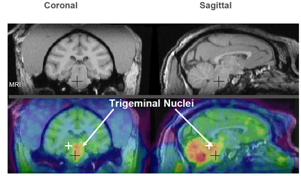
A collage of four MRI scans of the brain each showing a different view of the human brain. The first scan on the top left shows the left side of the image with the brain in the center. The brain is shown in black and white with a yellow square in the middle. The yellow square is likely the location of a tumor or a tumor in the brain. The image is labeled with the letters "A" "B" "C" "D" "E" "F" "G" "H" "J" "K" "L" "M" and "E". In the top right scan the brain appears to be in good condition with no visible signs of damage or damage. The top left and bottom right scans show the same brain but with a different color scheme. There are also several yellow squares scattered throughout the image. These squares are likely representing different areas of the tumor such as the cerebellum cerebrospinal cord and brainstorm. These areas are likely to be affected by the tumor or other abnormalities in the body. The images are arranged in a grid-like pattern with each square representing a different area. The background of each scan is black making the yellow squares stand out.
Description 
Type
Category
Source 1 of 4
-
Date
2018
Collection
-
Date
2018
Collection
-
Date
2018
Collection
-
Date
2018
Collection
We encourage you to view the image in the context of its source document(s) and cite the source(s) when using these images. However, to cite just this image alone, click the “Cite This Image” button and then paste the copied text.