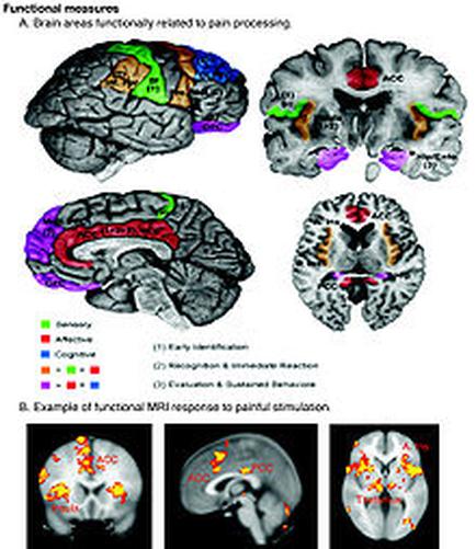
A collage of four different views of the brain each showing a different type of functional measures related to pain processing. The top left view shows the brain with different areas of the human brain including the cerebellum the cerebrospinal cord and the brainstorm. The top right view shows a normal brain with a red and green area in the center which is likely the location of the pain processing area. The brainstorm is colored in red indicating that the pain is caused by pain. In the bottom left view there is a normal MRI scan of the head and neck which shows the location where the pain occurs. The image also shows a comparison of the different areas with the red area representing pain and the green area representing inflammation. The red area represents pain while the orange area represents inflammation which may be caused by the pain in the brain. The text below the image explains that the image is an example of a functional MRI response to painful stimulation which can be seen in the top right corner of the image.
Description 
Type
Category
-
Date
2016
Collection
We encourage you to view the image in the context of its source document(s) and cite the source(s) when using these images. However, to cite just this image alone, click the “Cite This Image” button and then paste the copied text.