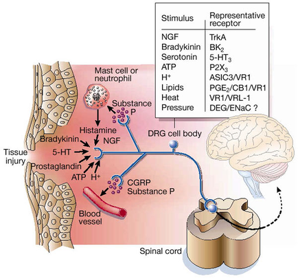
A diagram that shows the structure of a brain and its functions. On the left side of the image there is a tissue injury in the brain which is caused by a mast cell or neutrophil. The mast cell is responsible for the release of hormones and neurotransmitters in the body. On the right side there are two parts of the brain - the brainstorm and the spinal cord. The brainstorm is shown in the top right corner representing the representative receptor. The representative receptor is represented by a blue circle with the letters "NGF" and "Bradykinin" written on it. The blood vessel is shown as a red blood vessel which helps to regulate the flow of blood from the brain to the spinal column. There is also a diagram in the bottom right corner that shows how the blood vessel can be used to regulate blood pressure and regulate the blood flow. The diagram also shows the presence of the neurotransmitter which can be seen in the lower part of the body as well as the presence in the blood vessels.
Description 
Type
Category
Source 1 of 3
-
Date
2014
Collection
-
Date
2014
Collection
-
Date
2014
Collection
We encourage you to view the image in the context of its source document(s) and cite the source(s) when using these images. However, to cite just this image alone, click the “Cite This Image” button and then paste the copied text.