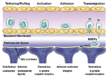
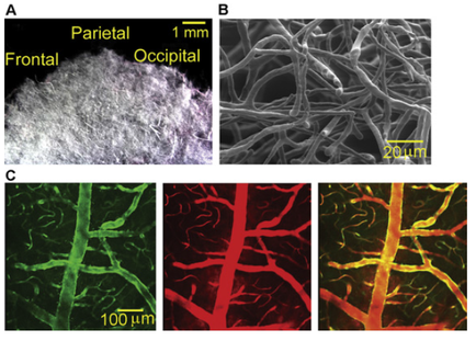
A collage of four different images. The first image on the top left shows a close-up of a parietal which is a type of nerve cell. The parietal is 1 mm in diameter and is located in the center of the image. It appears to be a microscopic view of the outermost part of the brain. In the top right image there is a black and white image of a group of neurons. The neurons are arranged in a radial pattern with some overlapping each other. The image is labeled with the letters "A" "B" "C" "D" "E" "F" "G" "H" "J" "K" "L" "M" and "E". Below the images there are four smaller images in the bottom left corner each showing a different color - green red and orange. The green image shows a tree-like structure while the red and orange images show a tree with red and yellow branches. The trees appear to be neurons and the image is taken from a top-down perspective.
Description 
Type
Category
-
Date
2015
Collection
We encourage you to view the image in the context of its source document(s) and cite the source(s) when using these images. However, to cite just this image alone, click the “Cite This Image” button and then paste the copied text.