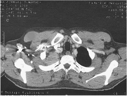
A black and white CT scan of a human skull. The skull appears to be in the middle of a cross-sectional view as there are several bones visible in the image. The bones are arranged in a circular pattern with the largest bone in the center and two smaller bones on either side. The largest bone on the left side of the image is the skull while the smaller bone is on the right side. The image also shows a small amount of blood vessels which are visible on the top left and bottom right sides of the skull. There are also several small structures including a large hole in the top right corner which could be a tumor or a tumor. The image is labeled with the names of the bones such as "R" "C" "D" "E" "F" "G" "H" "J" "K" and "L" indicating that it is an MRI scan. The scan is likely used to diagnose and treat a patient's heart attack or other medical condition.
Description 
Type
Category
Source 1 of 2
-
Date
2015
Collection
-
Date
2015
Collection
We encourage you to view the image in the context of its source document(s) and cite the source(s) when using these images. However, to cite just this image alone, click the “Cite This Image” button and then paste the copied text.