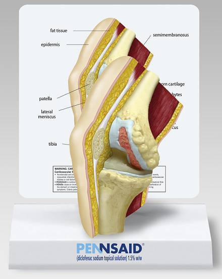
A photograph of a medical model showing a knee. It is a cross-section showing parts of the knee including the epidermis fat tissue patella lateral meniscus tibia semimembranosus and others. The model is sitting on a stand which appears to be made of white cardboard. The base of the stand has the Pennsaid logo.
Description
Category
-
Date
2010
Collection
We encourage you to view the image in the context of its source document(s) and cite the source(s) when using these images. However, to cite just this image alone, click the “Cite This Image” button and then paste the copied text.

